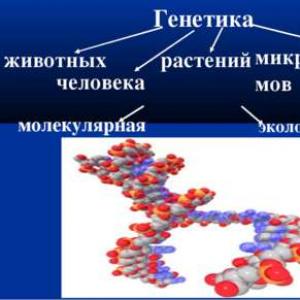
Features and methods of studying human genetics presentation. Biology presentation "methods of studying human genetics"
Cytogenetic method The cytogenetic method for studying human heredity is a microscopic analysis of chromosomes. Cytogenetics is a branch of genetics that studies the patterns of heredity and variability at the level of cells and subcellular structures, mainly chromosomes.

During the period of cell division at the metaphase stage, chromosomes have a clearer structure and are available for study. Usually, human peripheral blood leukocytes are examined, which are placed in a special nutrient medium, where they divide. Then preparations are prepared and the number and structure of chromosomes are analyzed.


Klinefelter's syndrome (47 XXY): the genetic essence of Klinefelter's syndrome is that instead of the normal male genotype with one X chromosome and one Y chromosome (46, XY), the patient will have one (or more) extra sex chromosome - X. The genotype of such a man will be (47, XXY).


Detection of many hereditary diseases is possible even before the birth of a child. The method of perinatal diagnosis consists in obtaining amniotic fluid, where the cells of the fetus are located, and in the subsequent biochemical and cytological determination of possible hereditary anomalies. This allows a diagnosis to be made early in pregnancy and a decision to continue or terminate.

1. Culturing blood cells on nutrient media 2. Stimulation of mitotic divisions 3. Adding colchicine to destroy the spindle filaments, stopping division at the metaphase stage 4. Treatment of cells with a hypotonic solution for free arrangement of chromosomes 5. Staining 6. Microscopy and photographing 7. Construction of an idiogram


"Hereditary syndromes and diseases" - Marfan syndrome, Albright's disease, dysostosis, otosclerosis, paroxysmal myoplegia, thalassemia, etc. Polygenic diseases (multifactorial). Genomic mutations Chromosomal mutations. Autosomal dominant diseases. Genomic mutations. Monogenic diseases. http://l.foto.radikal.ru/0612/08e0016d1d34.jpg. Polygenic diseases are not inherited according to Mendel's laws.
"Congenital diseases" - Disruptions. Most patients are missing one X chromosome. In fig. s-m Patau. Medical genetics. Private syndromology. Dysmorphology. In patients with profound mental retardation up to 1%, a 5p- deletion is found. Congenital malformations. Hereditary autosomal dominant disease.
"Human genetic diseases" - Mutations are hereditary changes in genetic material. Gene diseases and abnormalities. Hereditary diseases. Tell me what a man is. Indicate the possible genotypes of the parents. R: HSXs x HSU G: HS: HS: HS: U F1: HSHS: HSXs: HSU: HSU son - color blind. Pedigree chart. Mutations are hereditary, i.e. are steadily passed down from generation to generation.
"Cornelia De Lange Syndrome" -. From the side of the coordinating sphere - mimic the fall with intent while performing tests. Most often, the lag in intellectual development corresponds to mental retardation in the degree of imbecility. X-ray of the skull often reveals the phenomena of intracranial hypertension. IQ from 40 to 69. There was a passive fetal movement, breech presentation.
“Hereditary Human Diseases” - Ultrasound; Chorionic biopsy; Amniocetosis. What hereditary diseases do you know? In such families, there is a sharp increase in the frequency of hereditary diseases. Methods of prenatal (prenatal) diagnostics. About 2000 hereditary diseases and deformities are known. The nucleotide sequence of all human chromosomes has been deciphered.
"Chromosomal diseases" - A typical example of codominance is the inheritance of blood groups of the AB0 system in humans. Used precise quantitative methods to analyze data. Frequency of occurrence 1: 1000 Karyotype - 47, XXY, 48, XXXY, etc. Chromosomal. N / a: Phenylketonuria, Microcephaly, Ichthyosis, Progeria. Rules for the transfer of hereditary traits.
There are 30 presentations in total
Description of the presentation for individual slides:
1 slide
Slide Description:
Modern methods of studying a person The presentation was prepared by a student of grade 8 "A" of the Secondary School No. 50 Romanova Anastasia
2 slide
3 slide
Slide Description:
Audiometry Measurement of hearing acuity, i.e. the sensitivity of the auditory organ to sounds of different heights. It consists mainly in maintaining the lowest sound strength at which it is still heard. Three main methods are used: a study of hearing with speech, tuning forks, and an audiometer. The simplest and most accessible method is the study of hearing by speech. Its advantage is the ability to conduct an examination without special devices, in addition, this method corresponds to the main role of the auditory function - to serve as a means of verbal communication. Under normal conditions, hearing is considered normal when whispering speech is perceived at a distance of 6-7 meters. When using the equipment, the results of the study are entered on a special form: this audiogram gives an idea of the degree of hearing impairment and the localization of the lesion.
4 slide
Slide Description:
Biopsy An intravital excision of tissue or organs for examination under a microscope. It allows you to accurately determine the existing pathology, as well as diagnose clinically unclear and initial stages of neoplasm, and recognize various inflammatory phenomena.
5 slide
Slide Description:
Vectorcardiography Registration of the electrical activity of the heart using special devices - the vector of electrocardioscopes. Allows you to determine the change in the magnitude and direction of the electric field of the heart during the cardiac cycle. The method is a further development of electrocardiography. In the clinic, it is used to diagnose focal myocardial lesions, hypertrophy of the ventricles of the heart (especially in the early stages) and rhythm disturbances. Studies are carried out with the patient supine, placing electrodes on the surface of the chest. The resulting potential difference is recorded on the screen of the cathode-ray tube.
6 slide
Slide Description:
Cardiac catheterization Introduction of special catheters into the heart cavity through peripheral veins and arteries. It is used to diagnose complex heart defects, to clarify indications and contraindications for surgical treatment of a number of diseases of the heart, blood vessels and lungs, to identify and assess heart, coronary and pulmonary insufficiency. Catheterization does not require any special preparation of the patient. Usually it is carried out in the morning (on an empty stomach) in an X-ray operating room (with special equipment) by professionally trained doctors. The technique is based on the introduction of catheters into the heart through the aorta by puncture of the right femoral artery. After the study, patients need bed rest for the first day. Catheterization allows you to study the structure and function of all parts of the cardiovascular system. With its help, it is possible to determine the exact location and size of individual cavities of the heart and large vessels, identify defects in the septa of the heart, and also detect abnormal vascular discharge. Through the catheter, you can register blood pressure, electro- and phono-cardiogram, obtain blood samples from the heart and great vessels. It is also used for medicinal purposes for the administration of drugs.
7 slide
Slide Description:
Monitoring observation Is carried out for several hours or days with continuous registration of the state of the body. Monitoring is carried out over the pulse and respiration rate, the value of arterial and venous pressure, body temperature, electrocardiogram and other indicators. Usually, monitor observation is resorted to: 1) for immediate detection of conditions that threaten the patient's life, and for the provision of emergency assistance; 2) to register changes over a given time, for example, to fix extrasystoles.
8 slide
Slide Description:
Determination of eye pressure The aim of the study is to identify pathological changes in the tone of the eyeball. Both an increase and a decrease in intraocular pressure can impair the function of the eye and lead to severe, irreversible changes. The method is used to diagnose early glaucoma. Tonometers and elastotonometers are used to accurately determine intraocular pressure. The study is carried out with the patient lying down. After anesthesia of the eye with dicain solution, the doctor places the tonometer on the center of the cornea
9 slide
Slide Description:
Radioisotope diagnostics Recognition of pathological changes in the human body using radioactive compounds. It is based on registration and measurement of radiation from drugs introduced into the body. With their help, the work of organs and systems, metabolism, blood flow rate and other processes are studied. In radioisotope diagnostics, two methods are used: 1) The patient is injected with a radiopharmaceutical, followed by a study of its movement or unequal concentration in organs and tissues. 2) Labeled substances are added to the test tube with the blood to be examined, assessing their interaction. This is the like. screening test for early detection of various diseases in an unlimited number of people. Indications for radioisotope research are diseases of the endocrine glands, digestive organs, as well as bone, cardiovascular, hematopoietic systems, brain and spinal cord, lungs, excretory organs, and lymphatic apparatus. It is carried out not only with suspicion of some pathology or with a known disease, but also to clarify the degree of damage and assess the effectiveness of treatment. There are no contraindications to radioisotope research, there are only some restrictions. Comparison of radioisotope data, X-ray and ultrasound data is of great importance.
10 slide
Slide Description:
X-ray diagnostics Recognition of injuries and diseases of various organs and systems of a person on the basis of obtaining and analyzing their X-ray image. In this study, the X-ray beam, passing through the organs and tissues, is absorbed by them to an unequal extent and at the exit becomes non-uniform. Therefore, getting then on the screen or film, causes the effect of shadow exposure, consisting of light and dark parts of the body. At the dawn of radiology, the area of its application was only the respiratory system and the skeleton. Today the range is much wider: the gastrointestinal, biliary tract, kidneys, urinary system, blood and lymph vessels and other organs and systems. The main tasks of X-ray diagnostics: to establish whether the patient has any disease and to identify its distinctive features in order to differentiate with other pathological processes; accurately determine the location and extent of the lesion, the presence of complications; to assess the general condition of the patient. Organs and tissues of the body differ from each other in density and ability to x-ray transmission. So, well, bones and joints, lungs, heart are visible. When X-rays of the gastrointestinal tract, liver, kidneys, bronchi, vessels, the natural contrast of which is insufficient, resort to artificial, specially introducing into the body harmless radiopaque substances. These include barium sulfate, organic iodide compounds. They are taken orally (when the stomach is examined), injected into the bloodstream intravenously (with urography of the kidneys and urinary tract) or directly into the organ cavity (for example, with bronchography). The indications for X-ray examination are extremely broad. The choice of the optimal method is determined by the diagnostic task in each specific case. They usually start with fluoroscopy or radiography.
11 slide
Slide Description:
Rheographic research Rheography (literal translation: "rheo" - flow, flow and its graphic representation). A method for studying blood circulation, based on measuring the pulse wave caused by the resistance of the vessel wall when an electric current is passed. It is used in the diagnosis of various kinds of vascular disorders of the brain, extremities, lungs, heart, liver, etc. Rheography of the extremities is used in diseases of peripheral vessels, accompanied by changes in their tone, elasticity, narrowing or complete blockage of the arteries. The rheogram is recorded from symmetrical areas of both limbs, on which electrodes of the same area, 1020 mm wide, are applied. To find out the adaptive capabilities of the vascular system, tests with nitroglycerin, physical activity, and cold are used.
12 slide
Slide Description:
Thermography A method of recording infrared radiation from the surface of the human body. It is used in oncology for the differential diagnosis of tumors of the salivary and thyroid glands, bone diseases, cancer metastases in bones and soft tissues. The physiological basis of thermography is an increase in the intensity of thermal radiation over pathological foci due to increased blood supply and metabolic processes in them. A decrease in blood flow in tissues and organs is reflected by the "extinction" of their thermal field. The preparation of the patient provides for the exclusion of hormonal drugs, drugs that affect vascular tone, and the imposition of any ointments within ten days. Abdominal thermography is performed on an empty stomach. There are no contraindications, the study can be repeated many times. As an independent diagnostic method, it is rarely used; comparison with the data of the clinical and X-ray examination of the patient is mandatory.
13 slide
Slide Description:
Phonocardiography A method of recording sounds (tones and noises) arising from the activity of the heart and is used to assess its work and recognize violations, including valve defects. Phonocardiogram is recorded in a specially equipped isolated room where complete silence can be created. The doctor determines the points on the chest, from which the microphone is then recorded. The patient's position during recording is horizontal. The use of phonocardiography for dynamic monitoring of the patient's condition increases the reliability of diagnostic conclusions and makes it possible to assess the effectiveness of treatment.
14 slide
Slide Description:
Electrocardiography Registration of electrical phenomena occurring in the heart muscle when it is excited. Their graphic representation is called an electrocardiogram. To record an ECG, electrodes are applied to the limbs and chest, which are metal plates with sockets for connecting wire plugs. The electrocardiogram determines the frequency and rhythm of cardiac activity (duration, length, shape of teeth and intervals). Some pathological conditions are also analyzed, such as thickening of the walls of one or another part of the heart, heart rhythm disturbances. Possible diagnostics of angina pectoris, coronary heart disease, myocardial infarction, myocarditis, pericarditis. Some medications (cardiac glycosides, diuretics, cordarone, etc.) affect the readings of the electrocardiogram, which allows individual selection of medications for the patient's treatment. The advantages of the method - harmlessness and the possibility of using it in any conditions - contributed to its widespread introduction into practical medicine.
15 slide
Slide Description:
Electroencephalography A method of an electroencephalographic objective study of the functional state of the brain, based on the graphic registration of its biopotentials. The most widely used in solving the following problems: to establish the localization of the pathological focus in the brain, differential diagnosis of diseases of the central nervous system, to study the mechanisms of epilepsy and its early detection; to determine the effectiveness of the therapy and to assess reversible and irreversible changes in the brain. During the recording, electroencephalography, the examined person sits reclining in a special comfortable chair or, in serious condition, lies on a couch with a slightly raised headboard. Before the examination, the patient is warned that the recording procedure is harmless, painless, lasts no more than 20-25 minutes, that it is imperative to close your eyes and relax your muscles. Use tests with opening and closing the eyes, with irritation by light and sound. The electroencephalogram readings for any disease should be correlated with the data of the clinical examination.
16 slide
Slide Description:
Nuclear magnetic resonance Selective absorption of electromagnetic radiation by a substance. Using this method, it is possible to study the structure of various organs. The low energy of the used radiation significantly reduces the harmful effect on the body. The advantage of the method is its high sensitivity in the image of soft tissues, as well as high resolution, down to fractions of a millimeter. Allows you to get an image of the investigated organ in any section and reconstruct their volumetric images.
Slide 2
Slide 3
Lesson Objectives:
- Consider the features of the study of human genetics.
- Get acquainted with the basic methods of studying human genetics.
- Learn to use genealogical symbols when drawing up the pedigree of your family.
Slide 4
Lesson plan:
- Organizing time
- Goal setting, knowledge actualization:
- Learning new material
- Solving genetic problems
- Generalization and systematization of knowledge
- Test control of knowledge
- Defining and explaining homework, assigning marks.
Slide 5
Knowledge update:
What science deals with the study of heredity and variability?
- Genetics
Slide 6
Who is the founder of genetics?
Gregor Mendel
Slide 7
What object did Mendel use for his research? Why?
Slide 8
What method did Mendel use?
Slide 9
Can this method be used to study human genetics?
Slide 10
1. Difficulties in studying human genetics.
2 Methods of studying human genetics.
3. Genealogical method
4. Use of pedigrees in solving problems in genetics.
Plan for learning new material
Slide 11
Difficulties in studying human genetics:
1. Low fertility.
2. Slow generational change
Slide 12
3. The impossibility of staging special studies.
4. A large number of chromosomes and a complex gene structure.
Slide 13
Methods for studying human genetics.
Slide 14
Twin method
Based on the study of the development of traits in twins.
Allows:
1. With the greatest accuracy, find out the hereditary predisposition to certain diseases.
2. To establish the nature of the normal and disturbed nervous system.
3. It makes it possible to differentiate the role of the environment and genotype in the development of the phenotype.
Slide 15
Identical twins.
- They always belong to the same sex and show striking similarities to each other.
- They have the same genotype and the differences between them will be solely due to the influence of the environment.
Slide 16
Fraternal twins
- Can be of the same or different sex.
- Look like brothers and sisters.
- They are not twins.
- The differences between them are the result of heredity.
Slide 17
Cytogenetic method.
- Study of the karyotype of people.
- Allows you to identify changes in the chromosome set.
Slide 18
- Down syndrome is an example of a chromosomal disorder.
- The development of this disease is associated with trisomy of 21 pairs of autosomes - in the patient's cells there are 47 chromosomes instead of 46.
Slide 19
Biochemical method.
- Allows you to establish metabolic disorders
- Find out the hereditary predisposition to the disease and prevent the development of the disease in a timely manner.
Slide 20
Population method
On a mathematical basis, it determines the frequency of distribution of certain genes in the human population.
Slide 21
Genealogical method.
- Study of hereditary traits of a person by pedigree.
- Proposed at the end of the 19th century by F. Galton.
- The inheritance of diabetes, deafness, schizophrenia, blindness and other signs has been proven.
F. Galton
Slide 22
Terms and conventions used to define the pedigree:
- Genealogy is a pedigree.
- Pedigri - (fr.) Pedigree
- Sibs are brothers and sisters, descendants of the same parents.
- The proband is the owner of a hereditary trait.
- Inbreeding is a closely related cross.
Slide 23
Slide 24
Slide 25
Analysis of the pedigree.
1. Establishing whether a given symptom or disease is an isolated one in the family or there are several cases (family in nature). If a trait occurs several times in different generations, then we can assume that it has a hereditary nature.
2. Determination of the type of trait inheritance.
- To do this, analyze the pedigree, taking into account the following points:
- Is the trait under study found in all generations
- How many members of the pedigree have this trait?
- Whether the frequency of the trait is the same in both sexes.
- It is more common in persons of what sex.
- To persons of what gender the sign is transmitted from a sick father and a sick mother.
- Are there families in the pedigree in which both healthy parents gave birth to sick children or both sick parents gave birth to healthy children?
- What part of the offspring has an inherited trait in families where one of the parents is sick.
Slide 26
Solve the problem:
The proband is sick with congenital cataracts. He is married to a healthy woman and has a sick daughter and a healthy son. The proband's father is sick with cataracts, and his mother is healthy. The mother of the proband has a healthy sister and parents. The paternal grandfather is sick, and the grandmother is healthy. The proband has healthy aunt and uncle on his father's side. The uncle is married to a healthy woman and has three healthy sons. What is the probability of a proband's daughter having sick grandchildren in the family if she marries a man who is heterozygous for cataracts?
Slide 27
Pedigree to the problem
Slide 28
Sinkwine
Genetics
Medical, detailed
Studies, observes, helps
Studies the patterns of heredity and variability.
Slide 29
Homework
1. Compile the pedigree of your family or any fairy-tale character.
2. Solve the problem (task number 9 in the workbook)
3. Complete the research work (task number 10 in the workbook)
View all slides
Slide 1
Research methods of human genetics.
Biology teacher Lysenkova O.V.

Slide 2
Genealogical method.
Designed in 1865 by F. Galton. Tasks of the method: Determination of the hereditary nature of the trait. Determining the type of inheritance. Lets you learn about chained inheritance.

Slide 3
The study of the pedigree revealed that the mutation that causes hemophilia in royal families first arose in Queen Victoria in England.

Slide 4
Cytogenetic method.
Study of the structure and number of chromosomes.

Slide 5
Chromosomal diseases.
Down syndrome (trisomy on chromosome 21)

Slide 6
Down syndrome occurs as a result of a genetic abnormality. The signs of people with Down syndrome were first described in 1866 by the English physician John Langdon Down, whose name served as the name for this syndrome. The syndrome occurs due to the process of chromosome divergence during the formation of gametes, as a result of which the child receives an extra chromosome 21 from the mother (in 90% of cases) or from the father (in 10% of cases). Most patients with Down syndrome have three 21 chromosomes instead of two; in 58% of cases, the anomaly is associated with the presence of not a whole extra chromosome, but its fragments. Of the characteristic external signs of the syndrome, a flat face with slanted eyes, wide lips, and a wide flat tongue is noted. The head is round, a sloping narrow forehead, the auricles are reduced with an adherent lobe, the eyes have a spotted iris. The hair on the head is soft, sparse, straight with a low growth line on the neck. For people with Down syndrome, changes in the limbs are characteristic - shortening and expansion of the hands and feet. Incorrect growth of teeth, high palate, changes in the internal organs, especially the alimentary canal and heart.

Slide 7
Patau syndrome (trisomy 13)
Trisomy 13 was first described by Thomas Bartolini in 1657, but the chromosomal nature of the disease was established by Dr. Klaus Patau in 1960. The disease is named after him. Patau syndrome has also been described in tribes on a Pacific island. It was believed that these incidents were caused by radiation from atomic bomb tests. In England and Wales during 2008-09. 172 cases of Patau syndrome (trisomy 13) were diagnosed, of which 91% of diagnoses were made prenatally. Of which: 111 ended in abortion, 14 cases of stillbirth / miscarriage / fetal death, 30 results remained unknown and 17 children were born alive. More than 80% of children with Patau syndrome die within the first month of life.

Slide 8
DEVELOPMENTAL FAULTS Nervous system: - deviations of mental and motor development; - microcephaly; - holoproencephaly (violation of the formation of the cerebral hemispheres); - structural defects of the eyes, including microphthalmia, Peters' anomaly, cataract, colob, dysplasia or retinal detachment, sensory nystagmus, cork vision loss and optic nerve hypoplasia; - meningomyelocele (spinal defect) Musculoskeletal and cutaneous: - polydactyly ("extra fingers") - low-set and deformed auricles; - protruding heel; - deformation of the leg, the foot looks like a swing; - omphalocele (abdominal defect, umbilical hernia); - an abnormal appearance of the hand; - overlapping the thumb with the fingers; - congenital absence of skin (there are no areas of skin / hair); - cleft palate, cleft lip (cleft palate). Urogenital: - abnormal genitals; - kidney defects. Others: - heart defects (defect of the interventricular septum); - one umbilical artery.

Slide 9
Edwards syndrome (trisomy on chromosome 18)
Edwards syndrome was named after Dr. John Edward, who described the first cases in 1960 and recorded the pattern of symptoms. Most children with this pathology die at the stage of embryonic development, this happens in 60% of cases. The prevalence of Edwards syndrome is on average 1: 3000-1: 8000 cases. About 80 percent of the victims are women.

Slide 10
DEVELOPMENTAL FAULTS Low birth weight regardless of gestational age; Characteristic changes in the head: deformed small skull, small forehead and mouth, narrowed eyes, irregularly shaped ears, etc.; Micrognathia (defects of the upper and lower jaws). The shape of the face is distorted, the wrong taste is formed; "Wolf's mouth" and "cleft lip". Clefts are not found in all children; Multiple malformations of the cardiovascular, urinary and digestive systems; Violation of the reflex of sucking and swallowing (there are great difficulties with feeding); Malformations of the musculoskeletal system (clubfoot, fusion of toes); Almost complete lack of physical and mental development.

Slide 11
Sex chromosome trisomies.

Slide 12
Klinefelter's syndrome is a genetic disorder in males, which is based on a genetically determined deficiency of testosterone. It develops as a result of doubling in the developing fetus of one of the most important carriers of genetic information (sex chromosome). As a result of such violations in the sexual set of genetic information, female genes prevail over male genes, which determines the symptoms of the disease. Often it is not possible to make a diagnosis in a timely manner, which further leads to a more severe course of the disease. The disease leads to infertility, therefore it is not transmitted from generation to generation.

Slide 13
Twin method.
In 1876, F. Galton proposed using the method of analysis of twins to differentiate the role of heredity and the environment in the development of various traits in humans (he introduced the concept of "upbringing" and "nature").

Slide 14
Types of identical twins.

Slide 19
Siamese twins.
Siamese twins are identical twins that are not completely separated during embryonic development and have common body parts or internal organs. Usually, a fertilized egg divides on the sixth day after conception. Siamese twins are formed if the egg divides very late, 14-15 days after fertilization. By this time, the cells of the embryo have specialized in such a way that complete separation of twins in the womb becomes impossible. The chance of Siamese twins being born is about one in 200,000 births. About half of Siamese twins are born dead. The resulting infant survival rate is 5-25%. More often, Siamese twins are female (70-75% of cases).

Slide 20
Perhaps the most famous pair of twins were the Chinese Chang and Eng Bunker (1811-1874), who were born in Siam (present-day Thailand). For many years they toured with the circus under the nickname "Siamese Twins", thus securing this name for all such occasions. Chang and Eng had fused chest cartilages (the so-called xyphopagic twins). In modern conditions, they could easily be separated. They died in January 1874, when Chang was the first to die of pneumonia while Eng was asleep. Finding his brother dead, Eng passed away, although he was healthy.

Slide 23
Medical genetic counseling.
Purpose: Prevention of the birth of a child with severe hereditary diseases. They work in all major cities of Russia. STEPS: Studying the pedigree. Counseling: the doctor predicts the likelihood of having a sick child. An official conclusion, with a doctor's recommendation.

Slide 25
Prenatal diagnostics.
She uses ultrasound diagnostics (ultrasound), and surgical techniques and laboratory methods (cytogenetic, biochemical, molecular genetic). Prenatal diagnosis is extremely important in medical and genetic counseling, since it allows you to move from a probable to an unambiguous prediction of the child's health in families with genetic complications.

Slide 27
Literature.
Prikhodchenko N.N., Shkurat T.P. Fundamentals of Human Genetics: Uch. pos. Rostov n / a, "Phoenix", 1997. - 368 p. 2.http: //downsideup.org/ru/chto-takoe-sindrom-dauna 3.http: //vse-pro-geny.ru/ru_disease_7_Syndrom-Patau_%D0%A1%D0%B8%D0%BD%D0 % B4% D1% 80% D0% BE% D0% BC-% D0% 9F% D0% B0% D1% 82% D0% B0% D1% 83.html 4.http: //baby-calendar.ru/vrozhdennye -poroki / sindrom-edvardsa / 5.http: //lookmedbook.ru/disease/sindrom-klaynfeltera/male 6.http: //ru.wikipedia.org/wiki/%D0%A1%D0%B8%D0%B0 % D0% BC% D1% 81% D0% BA% D0% B8% D0% B5_% D0% B1% D0% BB% D0% B8% D0% B7% D0% BD% D0% B5% D1% 86% D1 % 8B 7.http: //anomalshina.ru/view_post.php? Id = 25